

Surgeries for Glaucoma

Glaucoma is a group of eye diseases that damage the optic nerve, which sends visual information to the brain. This condition generally affects peripheral (side) vision first, but can lead to blindness over time. In this article, we discuss the main surgeries for glaucoma.
The most common cause of, and currently the only approved treatable risk factor for, glaucoma is increased internal eye pressure, also called intraocular pressure (IOP). The eye constantly produces a fluid called aqueous humor. This fluid nourishes the eye and keeps it inflated, similar to the way that air inflates a balloon and gives it shape. Normally, this fluid drains through a circular canal that’s supported and held open by a structure called the trabecular meshwork.
In the most common types of glaucoma, this natural drainage system becomes blocked, causing fluid to build up, and pressure to increase. This higher pressure damages your optic nerve. If left untreated, glaucoma can lead to blindness, but if identified early and treated appropriately, vision loss can be slowed and sometimes even prevented.
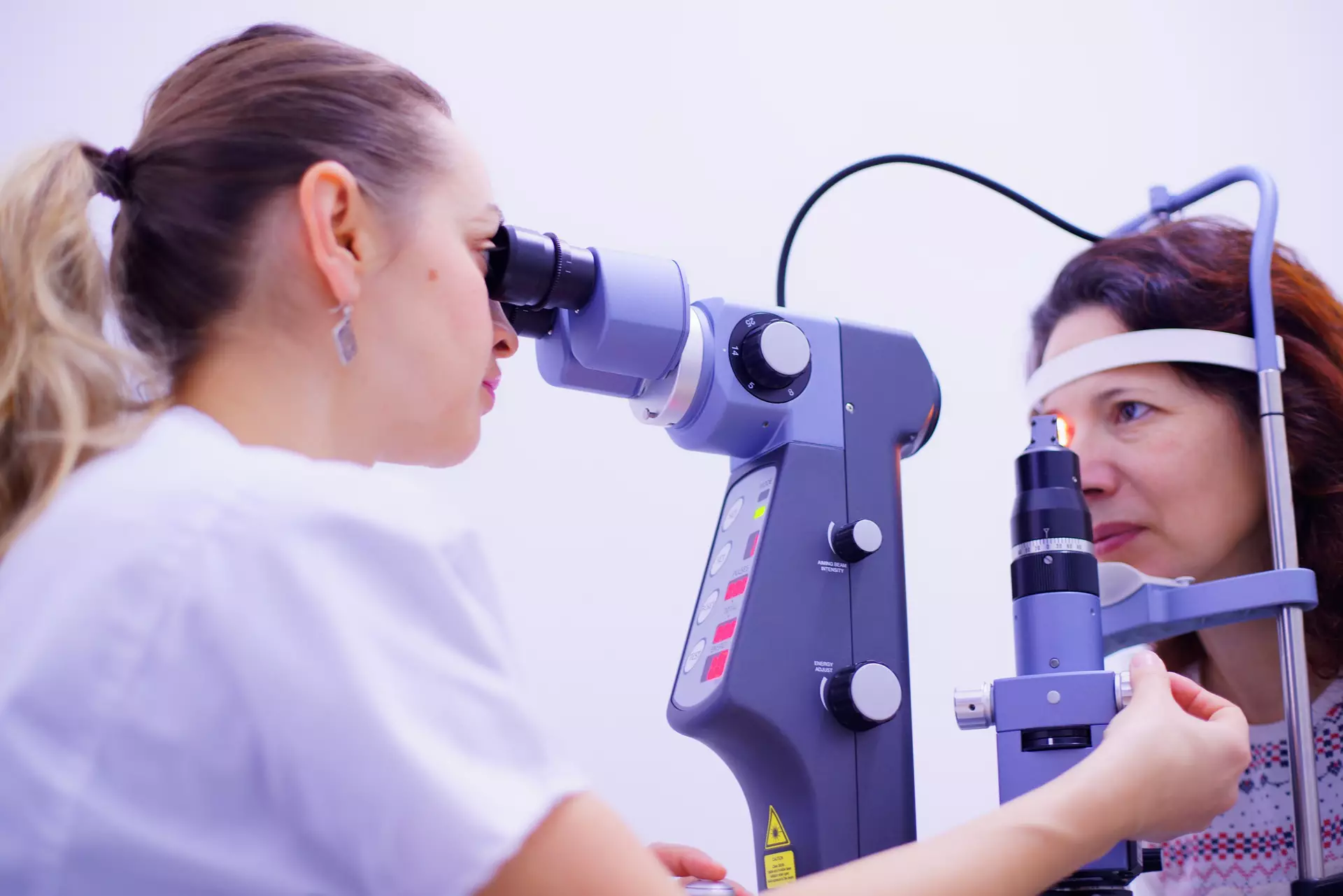
Glaucoma Surgery
(This image is from Pixabay.)
Why might surgery be recommended for glaucoma?
While medicated eye drops are the usual first-line treatment for glaucoma, sometimes surgery is necessary. Your ophthalmologist may recommend one of the many types of surgeries for glaucoma if:
- Other treatments have not been effective, or are unsafe to try
- Your glaucoma is severe or advanced enough to bypass milder treatments
- You have other medical conditions, eye-related or otherwise, that make surgery the best option
Fortunately, patients have several options. Here are several surgeries for glaucoma that your ophthalmologist may discuss with you.
Trabeculectomy
Trabeculectomy surgery is used to treat open-angle glaucoma. It takes around an hour or less to complete, and may be performed under local or general anesthesia, depending on whether you want to be awake (local) or asleep (asleep) during the procedure, and which method your doctor deems safest for you.
Your surgeon will administer anesthesia, then make an incision in your eye beneath your eyelid to create a channel, or “bleb,” to drain the excess fluid. Tiny stitches will hold the bleb open.
You’ll be discharged after the procedure, and will need to wear an eye patch until at least the next day when you return for a follow-up with your surgeon. Your stitches will be removed about two weeks after the surgery.
Tube shunt implant surgery
Your doctor may recommend implant, or tube shunt, surgery if you have neovascular or congenital glaucoma, though it can also be an option for people with other types of glaucoma.
This procedure takes approximately one to two hours, and involves implanting a tiny tube, also called a shunt, in the eye. The shunt lowers pressure in the eye by allowing extra fluid to drain. It’s typically done on an outpatient basis, using a local anesthetic and light sedation.
Minimally invasive glaucoma surgery (MIGS)
MIGS uses micro-incisions and miniscule devices to relieve pressure in the eye. This type of surgery often takes only minutes to complete, results in fewer complications, and has a faster recovery time than other types of glaucoma procedures.
MIGS is only used for mild cases of glaucoma, but it’s increasing in popularity and availability. Some experts, however, say they can’t be certain that MIGS procedures are as effective long-term as gold standards like trabeculectomy without more robust data.
The specialized equipment needed to perform MIGS also makes the procedure expensive, which can be a formidable barrier for patients without insurance.
Cyclophotocoagulation (CPC)
Cyclophotocoagulation is a laser treatment for glaucoma that targets the eye’s ciliary body, where fluid is produced. CPC works by reducing both pressure and fluid production.
You’ll remain awake during CPC. A local anesthetic is used to numb your eye, and the procedure takes just a few minutes. There are several different types of CPC, some being more invasive than others.
The corrections made by CPC are not necessarily permanent. The ciliary processes can return to overproducing fluid and increasing pressure.
Cataract surgery
Cataracts develop when a buildup of proteins clouds the lens of the eye, causing blurred and distorted vision. If caught early, cataract-related vision impairments can be managed with prescription eyeglasses, but advanced cataracts are best treated with surgery. Cataract surgery involves replacing the clouded eye lens with a clear, artificial one. While cataract surgery does not technically treat glaucoma, cataracts are a frequently co-occurring condition, and the surgery can help improve your vision.
Cataract surgery takes about 20 minutes, is performed under local anesthesia, and is usually painless, though you may experience some soreness, itchiness, and/or light sensitivity afterward. Your surgeon will likely give you an antibiotic and pressure-lowering eye drops to facilitate healing, along with a patch to protect your eye.
Prior to surgery, make sure your doctor knows about any underlying medical conditions you have, such as diabetes. Though complications from cataract surgery are rare, they can still occur.
Trabecular bypass stent surgery
Trabecular bypass stent surgery is usually only recommended for mild-to-moderate open-angle glaucoma. This outpatient procedure is typically performed during cataract surgery, and involves the implantation of a tiny, tube-like device through a small incision to provide an alternate path for outgoing fluid.
This surgery involves the same risks as cataract surgery alone, with the additional potential of stent blockage and/or dislocation.
Stent implantation might not be recommended for patients:
-
With angle-closure glaucoma
-
With conditions such as thyroid eye disease and Sturge-Weber syndrome
-
Who are juveniles
-
With chronic inflammatory disease
Canaloplasty
Canaloplasty is designed to enlarge the drainage canal with a microcatheter (small, thin tube) guided by a lighted fiber optic tip. Canaloplasty is performed by making a tiny incision in the eye.
The catheter is inserted into the canal and guided all the way through to remove blockages.
As the catheter is withdrawn, a sterile gel is injected to dilate the canal.
No devices are implanted in the eye, and the procedure may even reduce the need for glaucoma medication.
Potential risks and complications
As with any medical procedure, all surgeries for glaucoma can carry risks. Complications after surgery can include:
- Bleeding in the eye
- Cataracts
- Eye dryness
- Eye pain or discomfort
- Infection
- Low pressure in the eye
- Loss of vision
- Scar tissue formation
After surgery, it may take up to a few weeks for your eye to fully heal. Aftercare instructions will vary depending on the procedure performed.
Speak with your doctor about the benefits and risks of all your options to determine the best one for your situation and needs.
For more information on the pros, cons, side effects, and benefits of glaucoma surgery, download the Responsum for Glaucoma app.