The Four Types of Fibroids
The location of your fibroids in the uterus is what determines the type of fibroids you have. Understanding the different types of fibroids and knowing what kind you have will allow your doctor to create the most effective treatment plan for you.
Where do fibroids grow?
To understand the different types of uterine fibroids, you need to know that the uterus has different layers—three to be exact.
- The outer layer, or perimetrium
- The middle (and thickest) layer, or myometrium
- The inner layer that lines the uterus, or endometrium
What type of fibroids you have is defined by where, or on what layer, they’ve grown.
What are the different types of fibroids?
There are four main types of fibroids:
- Intramural fibroids
- Subserosal fibroids
- Pedunculated fibroids
- Submucosal fibroids
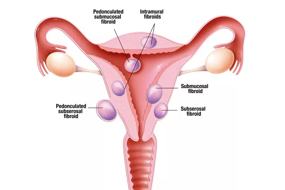
In this diagram from the National Women’s Health Network, you can see where each type of fibroid grows in the uterus.
Intramural fibroids
Intramural fibroids appear in the muscular wall of the uterus and are the most common type of fibroid. Due to the location of intramural fibroids, they may grow larger and can stretch the womb.
Subserosal fibroids
This type of fibroid grows outside of your uterus on the serous membrane (or serosa), which is the outer lining that all organs and internal body cavities have. Subserosal fibroids may grow big enough that they can make your womb look bigger on one side.
Pedunculated fibroids
Pedunculated fibroids form when a subserosal fibroid develops a stem. This stem turns into a slender base that can support a tumor. If this happens, this is where a pedunculated fibroid grows.
Submucosal fibroids
Submucosal fibroids grow in the myometrium, or middle layer of muscle in the uterus. This type of fibroids is not as common as the other three.
How can I find out what type of fibroids I have?
If you are experiencing any symptoms that could be a sign of uterine fibroids, schedule a doctor’s appointment to get tests performed and receive an official diagnosis. Normally, fibroids can be diagnosed through a pelvic exam. In a routine pelvic exam, your doctor can feel the size and shape of the uterus. If the uterus is enlarged or irregularly shaped, this is a strong indication of the presence of fibroids.

A pelvic exam is the first step in fibroids diagnosis. (This image is by Freepik.)
Further tests to confirm a fibroids diagnosis can include:
- Hysterosalpingogram (HSG) is a type of x-ray that allows you to see the inside of the uterus (uterine cavity). A device, called a cannula, is placed into the opening of the cervix and filled with a liquid that contains iodine to show contrast on the x-ray scan. This type of procedure can only identify fibroids that are inside the uterus (intramural fibroids).
- Transvaginal ultrasonography (TV US) is when an ultrasound wand, called a transducer, is inserted into the vagina. The transducer emits sound waves that bounce off of your organs and send images of the inside of your pelvis to a monitor. After this, your doctor or a technician will slowly turn off the transducer while it’s inside of you to get a complete picture of your organs.
- Saline infusion sonography (SIS), also referred to as sonohysterography (SHG), involves an ultrasound and sterile saline (saltwater) fluid, which work together to show the uterus and uterine lining. An ultrasound wand is placed in the vagina, followed by a catheter. The ultrasound exam continues while saline is injected through the catheter, which fills the uterus and allows the doctor to see an outline of the uterine walls and cavity. In doing so, this highlights abnormalities, such as fibroids.